I am really happy about finishing of my last blog for the year. Spending two weeks in Accident and Emergency (A&E) department has given me a very great insight about radiography. I have used this opportunity to see a lot of differences and similarities and acquired a great experience from my previous clinical placements.


This week’s blogging is going to be looking at how to accept personal responsibility for achieving the standards of professional behaviour as expressed in Health Professions Council Standards of Conduct Performances and Ethics (HPCSCPE).  As a student radiographer, this ‘’standards of conduct’’ has always been in my mind in order to ensure that I work for the best interest of the patients/ service users. Having this in mind gives me a great challenge on my day to day practice. Also each time I have a request card for any radiographic examination, I always bear in mind that as a health care professional, I am obliged to provide quality service to service user’s, I make sure I carry out my duties and responsibilities in a professional and ethical manner regardless of the patient age, gender, belief and religion, sexual orientation or physical ability.


According to Health and Care Professions Council, (2013) we have to’’ understand the need to respect, and so far as possible uphold, the rights, dignity, values and autonomy of every service user in maintaining health and wellbeing’’.


Areas whereby HPCSCPE is so much concerned about is respect to patients/ service user’s confidentiality.  While I was in every hospital/ radiography department, I have to follow the rules on ground in order to ensure that confidentiality is maintained at high level. HPCSCPE, stated that all patient information must be treated as confidential and use it for the purpose for which it was given. The procedure for handling patient information in every department is that after post processing of any request card, the request card has to be discarded to a special confidential waste bin, which is collected at the end of the day and dispose in a special way. I personally adhere to this rule throughout my stay in the department.  This is a good practice because it will help in handling confidential information relating to individuals at all times.

DH, (2012) on the other hand highlighted the importance of maintain confidentiality at all times. 

They put in place 8 principles of data protection described in the Data Protection Act 1998. One of the principle states that ‘’ everyone managing and handling personal information should understands that they are contractually responsible for following good data protection practice, is appropriately trained to do so and is appropriately supervised’’ This is a well reputable source and information gathered from them is up to date and it relates to everyday radiography practice, which I believe all staff have to adhere to these principle.


Being a student radiographer, it came to light to me that we have other duties and standards of conduct, performance and ethics we must comply with rather than taking x-rays.

  • We must keep high standards of personal conduct.
  • We must provide (any other relevant regulators) any important information about our conduct and competence.
  • We must keep our professional knowledge and skills up to date.
  • We must act within the limits of your knowledge, skills and experience and, if necessary, refer the matter to another practitioner.
  • We must effectively supervise tasks that we have asked other people to carry out.
  • We must get informed consent to give treatment (except in an emergency).
  • We must keep accurate records.
  • We must deal fairly and safely with the risks of infection.
  • We must limit our work or stop practising if our performance or judgement is affected by our health.
  • We must behave with honesty and integrity and make sure that our behaviour does not damage the public’s confidence in us or our profession.
  • We must communicate properly and effectively with service users and other practitioners.
  • We must make sure that any advertising you do is accurate (Health and Care Professions Council 2013).

I adhere to these rules in my day to day practice in the department by making sure that the right information is delivered to the patient, before, during and after their examination. I also do the same with fellow students and with post qualified radiographers. I see the reason why there is more emphasis on this because the word ‘’x-ray’’ is quite daunting to service users. The more we the radiographers work according to standard of conduct the more we maintain good reputation and the public will have confidence in our profession.


Researchers such as Williams and Berry, (1999) pointed out competency for newly qualified radiographers, which I considered vital to radiographer’s role. The study highlighted that radiographers should demonstrate knowledge of different modes of communication and when they should be utilized, including written, verbal and non-verbal, communicate effectively (basic skills, listening, observing, and interpreting) with: patients; relatives/carers; colleagues; other staff groups; wards; departments etc.


During this clinical placement, I have adopted a proactive approach to problem solving in all my clinical placement sites. This is one of the strength highlighted by all my supervising radiographers. A typical example whereby I have demonstrated a proactive approach to problem solving is how I have managed to contact one of the referrers (doctor) in A&E department.  A patient came to department on a trolley, unresponsive and in order for me to carry out his examination I needed to carry out positive identity check on him, but due to his unresponsiveness, I decided to use alternative means of identity check which is looking at his wrist band. I came to realise that he has no wrist band on, I had to inform my supervising radiographer and he ordered me to ring the doctor with his bleep number on the request card, so that they can send me a wrist band, which they did before I was able to x-ray the patient.


Other scenario whereby I have adopted a proactive approach to problem solving is by discovering a wrong address label on a request card, this mistake was done by the receptionist who attended the patient details and request on the computer before I picked up the request card. Also I had to inform my supervising radiographer and we spoke to the receptionist and she realised her mistake and did the correction with the right information before I could x-ray the patient.


Due to short of staff this week in the department, I was able to go to pain clinic in order to help the radiographer with some theatre work. Also, I have done a lot of mobile radiography while I was doing my A&E week; this has helped me to get all my mobile unassisted numbers for my mobile appraisal.

To top it up, I have managed to do my dependant patient appraisal this week, while I was going through the request card for the assessment I figured out that the referring clinician requested for a left elbow examination, because I considered checking for previous examination on (centricity) I realised that the card had a wrong request on it, instead of right elbow the referrer wrote down for left elbow examination. Also in this case, I adopted a proactive approach to problem solving by contacting the orthopaedics doctor and he made a change on the card before I can do the x-ray.


Reference List

Health and Care Professions Council (2013) Standards of proficiency.

Available From: http://www.hpcuk.org/assets/documents/10000DBDStandards_of_Proficiency_Radiographers.pdf

[Accessed on 13 March 2013].


Department of Health (2012) Data Protection Act 1998. Available From:  http://transparency.dh.gov.uk/dataprotection/  [Accessed on 13 March 2013].


Williams, P.L. and Berry, J.E. (1999) What is competence? A new model for diagnostic radiographers: Part 1. Radiography [online]. 5 (4), pp.221-235. [Accessed 13 March 2013].


With excitement of week nine on placement has made me to feel so confident to face the real radiography world. This week I have been working in Accident and Emergency department (A&E). I am happy with my ability in carrying out different radiographic examination, even though some of the A&E patient are quite challenging, to top it up I have been getting good comments from post qualified radiographer with regards to my adaptation of radiographic technique to suit patients physical/ psychological need .

During my time in A&E, I was opportune to observe one of the radiographer carrying out radiographic examination on a patient as a result of leg length measurements of the lower extremity.
Background reading from Golightly et al, (2007) defined leg length as a difference in lengths of the 2 legs. The study further highlighted that leg-length inequality may be caused by trauma, mild developmental abnormalities, onset in birth/childhood. It has been implicated as a cause of several conditions, such as trochanteric bursitis; knee pain; low back pain; osteoarthritis of the hip and knee; and running injuries, such as Achilles rupture etc.

In addition, the study also pointed out that different imaging modalities can be used to measure leg-length inequality, however radiography is the gold standard for measurement, other imaging modalities such as magnetic resonance imaging and computed tomography can be employed but they are expensive.

Slayton et al, (2012) in their study pointed out that other imaging technique can be employed for leg length examination as well using standard radiography include orthoroentogenogram, scanogram, and teleoroentgenogram. The only dilemma I have with this study is because the study was carried out in the U.S.A, so I believe their radiography practice is different from UK. However, the study made me to realise few limitations to all these techniques such as magnification error related to assessment of limb lengths, divergence of the x-ray beam etc.

The same study is also concerned about patient dose reduction in the utilisation of different radiographic technique. Due to recent advancement in technology, it came to light that microdose digital radiography is another form of computer- aided imaging that substantially reduces the radiation exposure to patients in comparison with conventional radiographic techniques. This technique works by using a vertical gantry, the patient stands in front of the x-ray assembly and remains stationary during the 20-second scanning process. According to the source, a patient receives an exposure of only 1 to 2 mrad during the scan; mrad is USA way of expressing x-ray exposure dose, looking at the dose recorded, it is well low dose compare to other radiographic techniques.

This examination was done on a normal computed radiography (CR) room, after processing the imaging cassettes, we realise that the image quality was too poor, with higher radiation dose recorded on the dose area product (DAP). As the superintendent he decided to re-examine the patient in the digital radiography (DR) room to ensure better image quality and to make sure diagnostic information are not missing. After the utilisation of digital scanning method for determining leg length. I evaluated the comparison on image quality, radiation dose on both systems.

By way of comparison, the DAP was ascertained from both systems. This measured value included the whole dose applied during the course of the examination. Collimation of the x-ray beam and tube filtration were taken into account.
Entrance dose recorded for this patient on a CR system for the whole leg length was 984 cGycm², with exposure of (90 kVp, and 60 mAs). And for the DR system entrance dose recorded for the same examination was 427 cGycm², with exposure of (120 kVp, and 60 mAs).This dose values shows that DR systems exhibits low radiation dose in comparison to a CR system.
A good image quality was produced using the DR rather than the CR system and that made it possible for the visualisation of correct area of interest such as the hip, knee and ankle joints, bony outlines was well defined, sharp trabecular pattern and adequate penetration with the exposure selected.

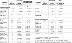
As part of my reflection during my academic session last year IDIS lecture on Radiobiology, I understand that Health Protection Agency, (2008) recommended patient dose information for different diagnostic examination. This highlights comparative radiation dose in the use of different radiographic technique. Please look at table below for more information.

DOSE TABLE

By doing this blog, I have been able to understand that different radiographic technique offers different radiation dose, due to maybe the body parts involve, i.e. Lumbar spine and abdomen requires a lot radiation in order to penetrate through the body tissues and acquire good images.

Kuhnley and Cueva, (2011) study pointed out that most of the increased exposure in the UK is due to CT scanning and nuclear imaging which require larger radiation doses than traditional x-rays. A chest x-ray, for example, delivers 0.02 mSv; while a chest CT delivers 8mSv see the table. This is 80 times as much. And that’s not counting the very common follow-up CT scans etc.

On the other hand, researchers such as Kuhnley and Cueva, (2011) estimated the potential risk of cancer from CT scans in 31,462 patients over 22 years. They highlighted that someone requires a CT or nuclear scan to treat or diagnose a medical condition; the benefits usually outweigh the risks. Still, if your clinician has ordered a CT, it’s reasonable to ask what difference the result will make in how individual condition is managed; for example, will it save an invasive procedure etc.

Also by doing this blog, I have utilised a wide range of different reference sources such as Health Protection Agency. This is well known independent UK organisation that was set up by the government in 2003 to protect the public from threats to their health from infectious diseases and environmental hazards. They provide advice and vital information to the general public to health professionals such as doctors and nurses, we the radiographers and to national and local government. Information gathered from them will serve as a guide in order ensure safe working environment and save life of individuals if we maintain the recommended radiation dose for different diagnostic procedure.

Other sources used such as Golightly, et al (2007) this source is not most recent, however it helped me to get a good background information about leg length and several conditions that can initiate discrepancy in leg length. Kuhnley and Cueva, (2011) source is most recent, it has helped me to rank different radiographic techniques in terms of the amount of radiation dose they can offer in comparison to traditional x-rays.

References:
Golightly, Y.M., Allen, K.D., Renner, J.B., Helmick, C.G., Salazar, A. and Jordan, J.M. (2007) Relationship of limb length inequality with radiographic knee and hip osteoarthritis. Osteoarthritis and Cartilage [online]. 15 (7), pp.824-829. [Accessed on 13 February 2013].

Health Protection Agency, (2013) Patient Dose information. Available From: http://www.hpa.org.uk/web/HPAweb&HPAwebStandard/HPAweb_C/119573382694 [Accessed on 09 March 2013].
Kuhnley, R. and Cueva, M. (2011) Learning About Cancer Has Brightened My Light: Cancer Education for Alaska Community Health Aides and Community Health Practitioners (CHA/Ps). Journal of Cancer Education [online]. 26 (3), pp.522-529. [Accessed on 13 February 2013].
Slayton, S., Williams, D.S. and Newman, G. (2012) Physical therapy services utilization and length of stay for obese patients following traumatic leg fracture. North Carolina Medical Journal [online]. 73 (1), pp.24-28. [Accessed on 13 February 2013].

My blog for this week is going to be focusing on my experience during the time I spent in minor injury unit (MIU). I worked with different radiographers, which gave me the opportunity to acquire more knowledge and learn new skills from post –qualified radiographers with about 35-40 years’ experience, at the end of the day I came to realise that, there are some advantages and disadvantages when working with different radiographers, from my personal experience, the advantages are more than the disadvantages. 

All the patients that came to the department throughout this week had minor injuries as the implies, most of the request came from the nurses in the same minor injuries department there and the examination request are mainly for upper and lower extremities with clinical indication such as crush injury, fall on outstretch hand (FOOSH), and minor trauma etc.

I have learnt a lot this week which gave me a good insight about radiography, and I will always bear this in mind as I progress in my career. First is the role of the radiographer in the environment of the healthcare team. As a radiographer, we have a responsibility under the ionising radiation (Medical Exposures) Regulation 2000 (IRMER), to justify any procedure involving ionising radiation, and to produce images of optimum diagnostic quality whilst keeping the radiation dose to patient as low as reasonably practicable –the ALARP principle (Department of Health,2007). This is due to the fact that ionising radiation has the potential to cause biological damage; the effect can be classified as somatic or genetic damage. 

Due to insufficient or poor knowledge of ionising radiation effect by other health care team, radiographers must make sure they maintain their role by making sure that any request card seeking for any medical examination utilising ionising radiation is properly validated, recent studies carried out by Mojiri, (2011) highlighted that even though there are some legislation and guidelines out there relevant to avoid risk of radiation incident some of the referrers’ still end up not meeting up with required criteria when referring patient for radiographic examination.  

In MIU department registered nurses, physiotherapist and podiatrists are permitted to ask for certain x-rays examination, i.e. radiographs of the upper and lower limbs.  While I was in the MIU department, I observed how the radiographers maintained their role among other healthcare team by making sure that any request is justified according to the agreed justification criteria in the department radiographic examination protocols and procedures.

Ionising radiation (Medical Exposures) Regulation 2000 (IRMER) in (North Bristol Trust protocol book) highlighted the duties of the radiographer. These duties are as follows.

  • Radiographers must positively identify patient before taken any x-ray examination.
  • Radiographers must ascertain pregnancy status of all female patients of childbearing age 12-55 years.
  • Other than the patient, everyone should stand behind protective screens during radiographic exposure.
  • X-ray room doors must be kept closed when making exposure.
  • Before irradiating any patient, the radiographer must make that it is the correct site of injury and must have correct clinical indication from the referrer.
  • Radiographer must make sure that gonad protection is used appropriately and should be suitable size and shape etc (North Bristol Trust, 2012).  

In addition, most of the patient who came to the MIU department had accompanied parents or relatives, one of the patients we x-rayed had a nurse and a healthcare assistant, during this time I experienced how the radiographer maintained his role within these other healthcare team, by making sure that adequate protection was utilised whilst the nurse assisted to immobilise the patient during the x-ray examination. This was a great experience to me.

Furthermore, it came to light to me while I was in the MIU department that radiographers role is not only just to comply to (IRMER), to justify any procedure involving ionising radiation, and to produce images of best diagnostic quality. Radiographers also must have the ability to work independently within a legal and ethical framework. Another area whereby a radiographer takes active participation in the department is by carrying out a routine quality assurance (QA) check on the x-ray equipment, my first day in MIU was on a Monday morning and the QA was due on that day, I observed the radiographer carrying out these checks.

This check is designed to ensure the best possible diagnosis at an acceptable radiation dose and with minimum cost. There are some guidelines stipulated by Institute of Physics and Engineering in Medicine, (2012) this is a well reputable organisation, which is external body that promote for the public benefit the progression of physics and engineering useful to medicine and biology and to advance public education in the field. Recommended checks according to (IPEM) for any routine QA by a radiographer are filtration, collimation, focal –spot size, alignment of the light beam diaphragm (Bushong, 2008).

A similar regulations to IPEM which is the Ionising Radiations Regulations 1999 (IRR’99). They deal with health & safety of those working with ionising radiation policed by the health & safety executive. Society and College of Radiographers, (2013) also recognise the need for this regulation because all radiographers are registered under them. Information gathered from this source is up-to-date, which is a professional body to all radiographers and it highlights some legal framework which employers and employees should adhere to under (IRR’99) to ensure patient and ionising radiation workers safety.

There are two main aims under (IRR’99). First, it is employer’s duties under the Regulations but should also be useful to others such as Radiation Protection Advisers (RPA), Health and Safety Officers, Radiation Protection Supervisors (RPS) and safety representatives. Secondly, the employees are legally obliged to comply with the Regulations and Local Rules and have a professional responsibility to rise with Employers, knowledge of procedures that are not fit for use.

A study carried out by Williams and Berry, (1999) which aimed at competency for newly qualified radiographers. Even though this study is not most recent, however it was published in diagnostic imaging journals, which I think the study should be considered as part of the radiographer’s role in the health care team. Examples of the statements.

  • Radiographers should demonstrate knowledge of different modes of communication and when they should be utilized, including written, verbal and non-verbal.
  • Radiographers should communicate effectively (basic skills, listening, observing, and interpreting) with: patients; relatives/carers; colleagues; other staff groups; wards; departments.
  • Liaise effectively with departmental staff, other departments, wards and clinicians etc.

This blog made me to learn a lot about radiographer’s role and how radiographers can work independently within a legal and ethical framework, such as IPEM, IRMER 2000, and IRR 99. Also I have recognised the fact that society and college of radiographers takes active contribution towards the wellbeing of its members to make sure that there is a mutual work relationship or agreement between employers and employees. In my future practice I will always bear this in mind and if radiographers keep to these roles and participate actively, it will enhance good quality care to the patient and our working environment will be safe.

Reference List.

Bushong, S.  (2008) Radiological Science for Technologists Physics, Biology and Protection. 9th ed. St. Louis: Mosby Elsevier.

Department of Health (2007) The Ionising Radiation (Medical Exposure) Regulations 2000. Available from: http://www.legislation.gov.uk/uksi/2000/1059/regulation/5/made [Accessed 28 February 2013].

Institute of Physics and Engineering in Medicine (2012) CODE OF PROFESSIONAL and ETHICAL CONDUCT. Available from: http://www.ipem.ac.uk/Portals/0/Images/Code%20of%20Conduct.pdf [Accessed 03 March 2013].

Mojiri, M. (2011) Awareness and attitude of radiographers towards radiation protection. Journal of Paramedical Sciences [online]. 2 (4), pp.2-6. [Accessed 28 February 2013].

North Bristol Trust (2012) Mobile and Theatre protocols. Available from: Protocol folder Southmead Hospital Room three. [Accessed 28 February 2013].

Society and College of Radiographers, (2013) Introduction. Available from: http://www.sor.org/learning/document-library/ionising-radiations-regulations-1999-irr99-guidance-booklet/introduction-0 [Accessed 03 March 2013].

Ionising Radiations Regulations (1999) HEALTH AND SAFETY. Available from: http://www.legislation.gov.uk/uksi/1999/3232/pdfs/uksi_19993232_en.pdf [Accessed 03 March 2013].

Williams, P.L. and Berry, J.E. (1999) What is competence? A new model for diagnostic radiographers: Part 1. Radiography [online]. 5 (4), pp.221-235. [Accessed 03 March 2013].

My blogging for this week is going to focus on the valuable insight gained from various modalities. I observed a  wide range of  ultrasound (US) examinations and they all involve using US gel, which allows the assessment of the  upper abdominal examinations (such as the kidneys, liver, and spleen etc) and testes.

Philip, (2009) describes US as a medical imaging modality which uses acoustic energy in order to create real-time images, which allow tissues to be localised and characterised. As ultrasound wave travels through the patient it will interact with layers between different tissues and this interaction will cause the wave to be attenuated.  This information so far has covered the principle behind using US, looking at the lecture covered on IDIS in 1st and 2nd year. 

While I was in US department, I witnessed a wide range of different US examinations such as assessment of the liver, gallbladder, bile duct, pancreas, kidneys, spleen and the associated vascular supply, abdominal aorta and testes etc.  I saw an interesting case where polycystic kidney disease was diagnosed (PKD). I devoted my time to find out more about this disease as this is something I have not heard of before.

PKD is described as an inherited disease, usually as an autosomal dominant trait. If one parent carries the gene, the children have a 50% chance of developing the disorder. The autosomal dominant PKD is said to occur both in children and adults, however it is much more common in adults, where symptoms often do not show up until middle age (Tortora and Derrickson, 2009).

From the diagnosis carried out on this young lady (26 year old) that came to the department, confirmed multiple clusters of cysts on her kidneys. A study carried out by Goilav, (2011) highlighted that the exact action that triggers cyst formation is unknown.

Tortora and Derrickson, (2009) confirms that in early stages of the disease, the cysts cause the kidney to swell, disrupting kidney function and leading to chronic high blood pressure and kidney infections. The cysts may cause the kidneys to increase production of erythropoietin, a hormone that stimulates production of red blood cells. This leads to too many red blood cells, rather than the anaemia seen in chronic kidney disease.

Even though US has more advantages of non-invasive procedure, patient’s acceptability, inexpensive, the lack of ionising radiation offer further rationale for selecting this imaging technique. However,  the only down side of this modality is that it cannot scan areas containing gas, due to issues with thermal effect, US cannot be used in obstetric scanning during development of the embryo and finally issues with cavitation, which means there might be risks of biological damage to tissue cells (Philip, 2009).

My time in Computed Tomography (CT) department made me to appreciate the clinical application of CT. CT works by measuring the attenuation of transmitted x-rays from the patient, which is converted to electrical signal and this electrical signal is amplified and analysed by the (DAS) data acquisition system (Carver and Carver, 2012).

While I was in the department, I observed a wide range of different CT examinations, which involves administration of contrast agent, and that allows the assessment of the abdomen, the chest and the head and neck etc.

One of the examination witnessed in the department was computed tomography pulmonary angiography (CTPA) looking at the pulmonary arteries. The clinical indication for this examination was for pulmonary embolism (PE).  This process involves administration of iodinated contrast agent, looking at the pulmonary arteries in order to identify any blockage of the main artery of the lung or one of its branches by a substance that has travelled from elsewhere in the body through the bloodstream.

After the examination, the report confirmed that the patient was suffering from PE. Among other modalities, Henzler et al,(2011) highlighted that the high spatial and temporal resolutions of multidetector-row CT (MDCT) has allowed CTPA to supersede perfusion scintigraphy and catheter angiography as the method of choice for imaging the pulmonary vasculature in suspected acute PE. The study confirmed that the current scanners visualize the pulmonary arteries down to the sixth-order branches; furthermore CTPA provides sensitive exclusion or specific diagnosis of acute PE. It is this definitive nature of test results that made CTPA the preferred modality for PE diagnosis.

In as much as CT is the most preferred choice in diagnosing such condition, which makes it more superficial to other modalities. However, the disadvantage of CT still remains that it offer high dose of ionising radiation to the patient (Mayo, 2009).

After my two days visit to the Magnetic Resonance Imaging (MRI) department, I feel like I have consolidated my knowledge from the theoretical aspect of my IDIS lecture last year on MRI applications. During my visit, I was able shadow the radiographers with the work they do.  At the same time I was excited because I had the opportunity to attach different coils on patient for each examination, I was able to help with the patient safety questionnaires before each examination.

Graham, Cloke & Vosper, (2011) describes MRI as the modality that uses a combination of magnetic field and electromagnetic radiation in the radiofrequency range in order to carry out diagnosis. I saw different diagnosis carried out using MRI.  The most interesting scan I saw was the upper abdomen scan (liver), which involved administration of contrast agent (Gadolinium). This patient previous report on ultrasound scan outlined coarse in echotexture on right lobe of the liver.

I was wondering why MRI would be a preferred modality choice in order to carry out further diagnosis on this patient instead of CT scan. According to De Toni et al, (2011) advantages of contrast-enhanced MRI for the liver include a high spatial resolution, better contrast sensitivity, better lesion detection and characterization than with CT. De Toni et al, (2011)suggested that few hepatic tumours, such as hemangiomas, carcinomas, and angiomas can be specifically diagnosed by their MRI appearance.

Reflecting to the lecture covered on MRI in my 1st year brings it to light to me that MRI does not make use of ionizing radiation compared to CT. In addition, in patients with allergies to contrast media. MRI contrast agents can safely be used in patients with allergies to iodinated contrast agents (De Toni et al, (2011).

These two days in MRI has helped me to see a lot of benefits around MRI such as excellent soft tissue detail, no use of ionising radiation etc. The main drawbacks of MRI include a long procedure time, and the need for the patient to hold their breath for longer periods and, it will not be a preferred choice of examination for claustrophobic patients.


Another two days spent in Nuclear Medicine (NM) department has been an eye opener to me, in fact I have considered furthering my career in NM.  This brings to mind what I have covered in IDIS lecture on NM, has giving me the opportunity to see the application in practice.

Mettler and Guiberteau, (2012) defines NM as an imaging modality which offers diagnostic images that provide information regarding the functional and physiological processes of a body system such as organ; tissue or bone, through the administration of a radionuclide attached to a pharmaceutical i.e. a radiopharmaceutical.

NM covers wide range of examination such as static renal scan i.e. ectopic kidney, and dynamic renal scan (renogram) etc (Major and Griffiths, 2009).

While I was in the department, I witnessed a wide range of different radionuclide examinations and they all involve administration of radiopharmaceutical product, which gives functional information.

One of the most common clinical indications is for cancer which ranges from different types of cancer scans. Can also be used to assess coronary heart diseases etc.  I had interest on a particular patient with prostate cancer and the query was to carry out a bone scan and the main reason for bone scan is to determine metastases of this disease. 

On his request there is an indication of high prostate specific antigen (PSA) and high Gleason score. According to PubMed Health, (2011) the PSA blood test is often done in order to screen men for prostate cancer. Due to the advent of PSA testing, some prostate cancers are now diagnosed early before they cause any symptoms. Other test such as CT scan or bone scan examination and biopsy is usually carried out in order to determine whether the cancer has spread.

The Gleason score on the other hand provides an effective measurement which helps to determine how severe the prostate cancer is, based on the appearance of the cancer cells when viewed under a microscope (PubMed Health, 2011). The radiographer I worked with made me to realise that the higher the Gleason score, the more likely the cancer is to have spread past the prostate. A typical example of the score is found below.

  • Scores 2 – 5: Low-grade prostate cancer.
  • Scores 6 – 7: Intermediate.
  • Scores 8 – 10: High-grade cancer. (PubMed Health, 2011).


The radiopharmaceuticals Tc-99m, hydroxymetylene diphosphonate (HMDP) is used while I was in the department is for bone scanning. Ramanlal et al, (2012) describes this product as one of the agent used for bone scanning. The study highlighted that metastasis in all cancers makes it difficult to control the disease spread even though some patient can respond to treatment.  The study also pointed out that bone is the most prone site for metastasis because of its physiological environment which enables tumour location and progress. Some common human cancers such as prostate cancer and breast cancer has high vulnerability (> 50%) to get metastasized at bone site in advanced stage and this is why this is why bone scanning is used to stage patients.


I have learnt more about prostate cancer and the part of the diagnostic process involved such as the Gleason score and PSA as well as using highly sophisticated equipment in order to diagnose patient or to determine the stages of the disease. This has made me feel good by doing work experience in nuclear medicine department, because I have more knowledge in addition to my background knowledge before my visit to the department.

By devoting my time doing this blog, I have good understanding of the theory principles which underpin diagnostic imaging modalities, I have seen the comparison in different modalities, the benefits and drawback involve in using all these different equipment.  The opportunity I had in order to see wider range of examination with all these imaging modalities, I believe all they said about what you see in theory is what is involve in practice.

My knowledge has been consolidated and I feel like am really prepared to face the real radiography world when I qualify. The most interesting issue during my modality weeks is that my supervisors highlighted that they (‘’were impress with my level of my knowledge and good under understanding for the stage of learning’’).

My clinical competencies mark shows above required standard for the time I spent in the department and no weakness or area of improvement indicated.

Reference List.

Carver, E. and Carver, B. (2012) Medical Imaging Techniques, Reflection and Evaluation. 2nd ed. Churchill Livingstone Elsevier.

De Toni, E.N., Gallmeier, E., Auernhammer, C.J. and Clevert, D.A. (2011) Contrast-enhanced ultrasound for surveillance of choroidal carcinoma patients: features of liver metastasis arising several years after treatment of the primary tumor. Case Reports in Oncology [online]. 4 (2), pp.336-342. [Accessed 24th February 2013].

Graham, T., Cloke, P., and Vosper, M. (2011) Principles and Applications of Radiological Physics. 6th ed. Churchill Livingstone Elsevier.

Goilav, B. (2011) Apoptosis in polycystic kidney disease. Biochimica Et Biophysica Acta (BBA) – Molecular Basis of Disease [online]. 1812 (10), pp.1272-1280. [Accessed 19 February 2013].

Henzler, T., Barraza Jr., J.M., Nance Jr., J.W., Costello, P., Krissak, R., Fink, C. and Schoepf, U.J. (2011) CT imaging of acute pulmonary embolism. Journal of Cardiovascular Computed Tomography [online]. 5 (1), pp.3-11. [Accessed 24 February 2013].

Mettler, F. and Guiberteau, M. (2012) Essentials of Nuclear Medicine Imaging. 6th ed. Philadelphia Elsevier Saunders.

Major, V. and Griffiths, M.  (2009) Nuclear Medicine. In: Easton, S (2009) An Introduction to Radiography. Philadelphia: Elsevier, pp.25-28.

Mayo, J.R. (2009) Radiation Dose in Cardiac CT. American Journal of Roentgenology (1976) [online]. 192 (3), pp.646-653. [Accessed 24th February 2013].

PubMed Health (2011) Prostate cancer. Available from: http://www.ncbi.nlm.nih.gov/pubmedhealth/PMH0001418/ [Accessed 24th February  2013].

Philip, R. (2009) Introduction to ultrasound. In: Easton, S (2009) An Introduction to Radiography. Philadelphia: Elsevier, pp.209-226.

Ramanlal Chaudhari, K., Kumar, A., Megraj Khandelwal, V.K., Ukawala, M., Manjappa, A.S., Mishra, A.K., Monkkonen, J. and Ramachandra Murthy, R.S. (2012) Bone metastasis targeting: A novel approach to reach bone using Zoledronate anchored PLGA nanoparticle as carrier system loaded with Docetaxel. Journal of Controlled Release [online]. 158 (3), pp.470-478. [Accessed 24th February 2013].

Tortora, G. J., and Derrickson, B. (2009) Principles of Anatomy and Physiology. 12th ed. John Wiley & Sons, Inc. U.S.A.


My time in placement this week has been spent on mobile x-rays examination, I was asked to focus more on mobile x-rays, because I needed to get my unassisted numbers for mobile x-rays which will enable me to carry out my mobile chest x-ray appraisal. During this time,  I realised that 70% of the mobile chest x-ray request that came to the department including the neonatal intensive care unit (NICU), were more on nasogastric (NGT) tube insertion position, lung collapse etc.

This made me to recall back on what I have learnt on IDIS module lecture last year on mobile /theatre and some clinical indications for mobile chest x-ray examination.  While I was working with the radiographers and due to issues that arise in the department regarding wrong NGT  insertion, this has  made me to devote my time to look at issues around  NGT such as patient care and risk involve if the tube is inserted to a wrong position (stomach).

According to Willcox, (2009) the purpose of NGT chest x-ray examination is to positively confirm that the exit hole of the (NGT) is in the right point within the gastrointestinal tract. When the nasoenteric tube is used the main aim is to make sure that the tip of the tube past the pylorus into the jejunum.  If this tube is placed properly, it helps to delivers nutrition/therapeutic agents directly into the jejunum when the patients are unable to feed through their mouth. Whereby the NGT is malpositioned the outcomes could lead to death.  However, the study highlighted that x-ray confirmation of NGT position is not practised in some hospitals; rather the position of the tip of the NGT is assessed by drawing back gastric contents and testing with pH paper.

In order to reduce the harm/death caused by misplaced NGT, National Patient Safety Agency, (2011) put in place the appropriate testing that needs to be carried out in order to make sure that the NGT are in the right position.

Method that should be:

  • Measurement of the pH of aspirate using pH indicator strips/paper.
  • Radiographic examination.


Methods that should NOT be used:

  • Auscultation of air insufflate through the feeding tube (‘whoosh’ test).
  • Testing the acidity/alkalinity of aspirate using blue litmus paper.
  • Interpreting absence of respiratory distress as an indicator of correct positioning.
  • Monitoring bubbling at the end of the tube.
  • Observing the appearance of feeding tube aspirate.

Even though National Patient Safety Agency (NPSA) recommended two methods to be used in order to make sure that NGT is placed in the right place, study carried out by Lemyze, (2010) pointed out that there are some limitations with pH testing and radiographic examination.

Limitation with the pH testing method is that the stomach pH can be affected by medication such as proton pump inhibitors, apart from ranitidine and frequency of feeds (Willcox, 2009). Furthermore, gaining aspirate from the feeding tube can be very hard, more especially when using fine bore tubes.

Multiple reports of x-rays being misinterpreted by radiologist is highlighted as another limitation. However, the most accurate method for confirming NGT placement is chest x-ray examination (Lemyze, 2010).


From this week’s placement I have understand the more reason why radiographic examination is used ‘routinely’ for chest x-ray examination for NGT, also I have seen the reason why room three local protocol recommends for particular groups of patients, i.e., those on intensive care units and neonates requires a mobile chest-ray examination because such examination is too sensitive in order to avoid more risks and patient death.


Reference List.

National Patient Safety Agency (2011). Reducing the harm caused by misplaced nasogastric feeding tubes in adults, children and infants. Available from: http://www.nrls.npsa.nhs.uk/EasySiteWeb/getresource.axd?AssetID=129697&. [Accessed 15 February 2013]

Lemyze, M. (2010) The placement of nasogastric tubes.(Five things to know about …). CMAJ.Canadian Medical Association Journal [online]. 182 (8), pp.802. [Accessed 15 February 2013]

Willcox, J. (2009) Nasogastric tube insertion. The Foundation Years [online]. 5, Supplement (0), pp.13-15. [Accessed 15 February 2013]

This week’s blog is going to be focusing on my theatre experience and  reflection of what I have learnt in theory in comparison to how things are done in practice. My first hour in the theatre was a confronting new environment. During my time in the theatre, I encouraged myself and felt like I was equipped with the background knowledge from the lecture I had on mobiles and theatre on IDIS.

While I was in the theatre, I figured out some strategies for dealing with different problems which could occur when carrying out operating theatre techniques.  Drafke and Nakayama, (2000) pointed out that theatre experience can be very pleasant or very difficult. I realised that in order to have a pleasant experience in the theatre it will be a good idea for a radiographer or as a student to be a good communicator with the theatre staff; this could involve such things as knowing their names, roles, and rules etc.  

As I could remember from IDIS lecture, I was advised to be aware of operating theatre rules, by keeping to this rules it will help make things much easier. These rules are present in operating theatre folders of protocols. These covers aspects of theatre procedures such as attire, infection control, occupational health and safety, patient safety etc (North Bristol Trust, 2012).  Because we the radiographers are just on a few hours  mission in this theatre, we are not  probably  be expected to study these protocols, but the few that affect us will need to be known and it will be a good practice to know more as additional information.

From my experience I gathered that as soon as the surgeon breaks the patient’s skin, the patient is at risk of acquiring infection. One of the goals of the operating theatre staff is to prevent the patient developing a significant post-operation infection (North Bristol Trust, 2012).  This will mean that high level of care should be taken to avoid touching anything or anyone that is sterile. This will mean not standing too close to a staff member who is gowned and gloved. It will also require us to point out to others in the operating theatre when they are at risk of breaching a sterile field, or indeed, when they have breached a sterile field.

Furthermore, extra care has to be maintained particularly when you pushing the image intensifier over a patient in order to ensure that you do not touch the sterile handle attached to the operating light. The key point for the radiographers is to  know  the image intensifier in and out, now I see the reason why some radiographers allows us the student to have a feel of handling the image intensifier, the radiographer I worked with the gave me the opportunity to carry out some task in the theatre i.e. pressing the exposure button etc.

However, some radiographers don’t really allow students to do much in the theatre and I see the reason for that, i.e. in major cases, there is no time in the theatre for radiographers to be teaching student due to the fact that the surgeons wants the image as soon as possible in order to avoid waste of time, that left me with a question to ask, WHEN DO WE HAVE TIME TO PRACTICE MORE ON THEATRE WORK? Because as a newly qualified radiographer, the most common job for us in the department is theatre work.

Also I was opportune to do some post-processing, by sending of the images acquired to Picture Archiving and Communications System (PACS), which I really like because it gives me the organisation skills in order to do things in a proper manner from selectecting the saved images for the surgery, sending it off to PACS, doing all the paper work- recording dose, and time etc.

According to Dunmall, (2009) identify the patient for each radiographic procedure is very crucial. The most common method for identifying the patient is from the name on the paperwork in the theatre. When the patent has been anasthestetised, it will be difficult to obtain identity check, alternative means to identify this patient is to look on wrist band and match it to what is on the request card/notes in theatre.  

Radiographers checks to be made in the theatre for patient undergoing any surgery. This involves information such as, patient personal identification (name, date of birth, and address), pregnancy status, mandatory requirement under, Ionisation Radiation Medical Exposure Regulations 2000 IR (ME) R. Clinical indication- this is information related to the area and mechanism of injury, investigation required- this is information relating to imaging procedure and operator’s details (Dunmall, 2009).

All this checks are made in order to comply with (IRMER). Messer, (2009) highlighted that it is the duty as a radiation worker  to carefully  minimize radiation exposure whenever possible and thus to apply the “As-Low-As-Reasonably Achievable”(ALARA) principle in order to ensure the lowest possible radiation dose to the patient and staff. As a radiographer we are obliged to abide by this regulation.

In addition, I understand that as a radiographer, we are meant to be aware of the procedures, because, there will be a variety of procedures that we will be expected to assist with. Also I realise that, it is better to spend 5 minutes at the beginning of an operation covering the image intensifier than to spend 20 minutes at the end of an operation cleaning off bodily fluid. Setting up the monitor and control panel prior to each examination is a useful idea, because it saves time.  Again, securing the mains cord has advantages. It will help hold cable in place in order to avoid damage to the image intensifier, even if someone trips over the cord.

I experienced a lot during my time in the theatre; also I got a lot of tips from the radiographers I worked with which I think will be beneficial to me once I qualified.  I was advised to go to the toilet before commencing any theatre procedure as one can never be sure how long an operation will take, check that the surgeon is operating on the correct side, always keep an eye on the surgeon’s eyes when screening, it is a radiation protection issue, more especially dose of radiation to upper extremities, studies conducted by   Back et al, (2005) have shown higher doses of radiation reaching the hands of the surgeons, than other parts of the body. By keeping an eye on the surgeon, it will help the radiographer to understand what they are doing at every stage of the operation and this help whether the surgeon have simply forgotten to ask the radiographer to stop screening.

I was advised to be extra careful when moving the C-arm for some many reasons, i.e. to avoid causing further damage to the patient, and staff which might cause obstruction to the entire surgery.  Distance, shielding and time, were also highlighted to me in order maintain the three basic principles of radiation protection (Bushong, 2008).

My experience this week in the theatre has made me to realise that as a radiographer, we have to be able to have full understanding of theatre work, because it is very embarrassing if a qualified radiographer don’t know how to perform a task on the image intensifier.  I realised that good communication skills will suggest to the surgeon that you deserve his or her confidence in your abilities. I learnt that it will be adviser able to seek for the surgeon’s guidance if unfamiliar with the procedure, by asking questions such as which side would you like me to come in from? Do you want the image intensifier to go under the table or over the table for the lateral? Having all this in mind I have the confidence to face my next week in the theatre alongside my mobile radiography.

Reference

Back, D.L., Hilton, A.I., Briggs, T.W.R., Scott, J., Burns, M. and Warren, P. (2005) Radiation protection for your hands. Injury [online]. 36 (12), pp.1416-1420. [Accessed 7 February 2013].

Bushong, S.  (2008) Radiological Science for Technologists Physics, Biology and Protection. 9th ed. St. Louis: Mosby Elsevier.

Dunmall, K. (2009) Clinical skills for preparation of the patient and clinical environment. In: Easton, S (2009) An Introduction to Radiography. Philadelphia: Churchill Livingstone, pp 53-65.

Drafke, M. and Nakayama, H. (2000) Trauma and Mobile Radiography. 2nd ed. Philadelphia.

Messer, S. (2009) The law at work. In: Easton, S (2009) An Introduction to Radiography. Philadelphia: Churchill Livingstone, pp 11-21.

North Bristol Trust (2012) Mobile and Theatre protocols. Available from: Protocol folder Southmead Hospital Room three. [Accessed 7 February 2013].

I am so excited that placement is on the 4th week now and that made me to realise that I have few months left to go into my final year that means very soon, I will become a qualified radiographer. Having all this in mind has made me to work so hard in order to achieve my dream despite the fact that I have to set my alarm to go off at 6:30am Monday –Friday, in order to get ready and be on placement site by 8:30am. I look forward to placement every day because I learn a lot from it and also it helps to amalgamate my knowledge with all the theoretical information delivered to us during lectures last year in SIDI and IDIS module.

Like I said I always learn new things every day on placement and I  also love the patient care side of radiography, it gives me the zeal when dealing with the patients in order to do all the necessary check such as ID check before carrying x-ray examination . This 4th week blog is going to be looking at one of the knee projection called ‘’Rosenberg’s view’’, which I came across when I did my 1st week at the minor injury unit (MIU).

According to Wiki radiography, (2011) the maximum stresses acquired in the knee joint occur between 30° and 60° of flexion, by carrying out standard anterior posterior (AP) weight-bearing radiographs on extension makes it  inappropriate to evaluate cartilage loss as it can be indicated by narrowing of space.

The Rosenberg view is best described as a 45-degree flexion, poster anterior, weight-bearing view of the knee with the patellae touching the image receptor. By carrying out this particular projection it creates a good joint space and narrowing, which makes this view more sensitive and specific to joint space rather than the conventional extension weight-bearing anterior posterior views, and is useful for the assessment of knees with early degenerative change (Wiki radiography, 2010).

Information in the box below describes how this examination can be carried out.

Name of projection.

Knee – PA Weight bearing   (Rosenberg’s) 

Area Covered.

Distal femur, proximal tibia and fibula, bilateral tibiofemoral joint   spaces under the effects of gravity.

Pathology shown.

Joint space narrowing due to cartilage degeneration or other knee   joint pathologies.

IR Size & Orientation.

35 x 43cm Landscape (CR and DR).

Exposure.

66 kVp 12 mAs.

FFD / SID.

100cm.

Central Ray.  

CR perpendicular to IR.

Centring point.

Directed to a midpoint between the knee joints at a level of the   patella apices.

Collimation.

Collimate to bilateral knee joint region include distal femur and   proximal tibia / fibula to see alignment of joints.

Marker orientation.

PA marker.

Shielding Gonadal.

Check according to department’s policy guidelines.

Patient Positioning.

  •   Patient should be standing.
  •   Patient facing the upright bucky.
  •   Position feet straight ahead with weight   evenly distributed on both feet support with handles if needed.
  •   Flex knees 45° with both patellae touching the   IR.
  •   CR perpendicular to IR, directed to a midpoint   between the knee joints at a level of the patella apices (Wiki radiography,   2010).


Study carried out by Fontboté et al, (2008) suggest that’’ Rosenberg projection should be included in the initial study of any patient with suspicion of knee osteoarthritis’’. Knee osteoarthritis is the most common disease in the UK which affect 8 million people which could be male or female above the age of 50years (Arthritis Research UK, 2013). It is important to carry out early diagnosis in to enable early treatment and eventually delay its progression.

This study compared the diagnostic yield for osteoarthritis of two projections in knee x-ray examination. They evaluated 44 knees with conventional (AP weight-bearing, in full extension) and Rosenberg (PA weight-bearing in 45 degrees of flexion) x- ray projections in 32 patients (24 women and 8 men), aged 26 to 78 years. All patients were symptomatic at the time of evaluation, with a suspicion of knee osteoarthritis. Both projections were analysed and compared.

The study concluded that Rosenberg projection is more sensitive to detect knee joint space narrowing as compared with conventional AP weight-bearing projection, at least in the medial compartment of the knee. This makes it vital for department to consider this projection as part of their protocol.

By doing this blog on Rosenberg view with regards to my PODI lecture in 1st year, I know see the reason why this projection is recommended because it allows true visualisation of knee  intercondylar eminences, all the condyles of femur (lateral and medial) etc.  Because of the anatomical location and the complex biomechanics of the knee, osteoarthritis changes are commonly found in the knee and radiographic examination like this should be undertaken if surgery is being considered.  

Reference List.

Arthritis Research UK (2013) What is osteoarthritis? Available from: http://www.arthritisresearchuk.org/arthritis-information/conditions/osteoarthritis/what-is-osteoarthritis.aspx [Accessed 31 January 2013].

Fontboté R C, Nemtala U F, Contreras O O, Guerrero R. (2008) Rosenberg projection for the radiological diagnosis of knee osteoarthritis. PubMed [Online]. 49 (4), pp.261-269. [Accessed 31 January 2013].

Wikiradiography (2011) Knee – Rosenberg’s. Available from: http://www.wikiradiography.com/page/Knee+-+Rosenbergs   [Accessed 31 January 2013].

My 2nd week on clinical placement blog will be looking at continuing professional development (CPD). As a registered member of the society college of radiographers, I do receive monthly journal of diagnostic imaging and radiation therapy and synergy news etc. As I read through all the information in these journals and synergy news, I came to understand that qualified radiographer, we will be taking part in doing (CPD) as part of a member of the health and care professional council (HCPC).
What is CPD? HCPC, (2012) defines continuing professional development(CPD) as ‘’a range of learning activities through which health and care professionals maintain and develop throughout their career to ensure that they retain their capacity to practice safely, effectively and legally within their evolving scope of practice’’.
From my own understanding, I gathered that registrants must undertake CPD to stay registered with HCPC and they set standards which members must follow in order to complete their CPD portfolio.
• Registrants should maintain a continuous with up-to-date and accurate record of their CPD activities.
• Every HCPC member should show that their CPD activity includes a mixture of learning activities relevant to practice currently or in future.
• Must ensure that their CPD has contributed to the quality of their practice and service delivery.
• Must ensure that their CPD benefits the service user.
• If CPD is requested by HCPC, registrants must present a written profile (which must be their own work and supported by evidence) explaining how they have met the standards for CPD.

In order to ensure that professionals maintain their CPD portfolios and up to date, HCPC randomly check the CPD of individuals when they renew their registration, people that are chosen must submit their CPD portfolio which has to show how their CPD meets standards.

Society of Radiographers, (2013) in the other hand support and encourage all members to engage in CPD and they also provides different resources in order to support their members and I believe that was what caught my interest in looking at CPD and issues around it.
CPD is structured in a way that every radiographer records their activities in a reflective manner, also reflective practice is said to be a vital tool in practice based professional in the sense that people will learn from their own experience.

I think it will be a nice idea if I should blog on this CPD, in order to make other fellow student be aware of the future. Also as a member of the society, I think I should utilise any opportunity that comes out from this monthly information from the society and that will keep me up to date with what is going on in the society, which will benefit me and other students from blogging on issues like this.

Reference List.
Health and care professional council (2012) Continuing professional development. Available from: http://www.hpc-uk.org/registrants/cpd/ [Accessed 16 January 2013]
Society of Radiographers (2013) Continuing professional development (CPD). Available from: https://www.sor.org/learning/cpd [Accessed 16 January 2013]

My blog for this week is going to be looking at contrast agent’s adverse reaction, in addition to what I have learnt from IDIS module lecture. During this week’s clinical placement, I was fortunate enough to be in intravenous based contrast studies department specifically dealing with computed tomography (CT) work, observing and assisting the radiographers as they carry out different radiographic examinations.
Thomsen, (2011) highlighted that different contrast agents has been used in order to ensure improvement on vascular imaging and adverse reaction occur due to administration of these contrast agents. The study pointed out that adverse reaction could be minor physiological disturbances to life threating condition. Due issues around contrast agents and the adverse reactions, I decided to look at the chemistry of iodinated contrast agent and other issues such as osmolality, viscosity and ionicity.
My findings, Caver and Carver, (2012) describe contrast agents as water soluble organic preparation in which molecules of iodine are the opaque agent. The compound contain iodine atoms, with atomic number 53, which bonds to a carrier molecule, and it helps to hold the iodine in stable compound which then makes it possible to be delivered to the organ under investigation.
Contrast agents are chemically modified from 2, 4, 6-triiodinated benzene ring. The carbon atoms on a benzene ring are numbered clockwise from 1 to 6. Benzoic acid is produced by introducing an acid group at position 1 on the benzene ring. This acid group allows the salts to be formed and it influences water solubility 2, 4, 6-triiodobenzoic acid which is obtained by introducing iodine atoms at positions 2, 4, and 6 on the ring.
The reason why iodine is used is because it has three important properties which are vital for the production of contrast agent: high-contrast density, firm binding to the benzene molecule, and low toxicity (Singh and Daftary, 2008).
According to Singh and Daftary,(2008) osmolality, viscosity and ionicity plays important role in the development of contrast reactions, because of the chemical properties of contrast agents, which makes it thicker (viscosity) and having greater osmolality (more molecules per kilogram of water) than blood, plasma, or cerebrospinal fluid.
Ionicity involves molecule break up, which is classified into positively charged+ and a negatively charged_, resulting in more molecules per kilogram of water and therefore increasing osmolality. Whereby, non-ionic agents do not have this property and hence are less osmolar.
The iodine contrast based compound are classified into four groups (Caver and Carver, 2012) known as ionic monomers, ionic dimers, non-ionic monomers, non-ionic dimers. Typically, 3 iodine atoms are delivered with each benzene ring of a contrast medium. If a contrast molecule contains only 1 benzene ring, it is called a monomer. To deliver more iodine with each molecule of contrast, 2 benzene rings may be combined to produce a dimer. This molecule would deliver 6 iodine atoms with each molecule.

Mechanism of action of iodine in a solution, “ionic monomers break up into their anion and cation components”, which allows increase in osmolality, and delivers 3 iodine atoms (a 2:3 ratio of osmolar particles to iodine), whereby ionic dimers usually delivers 2 ionic components per 6 iodine atoms (ratio, 1:3). In contrast” non-ionic monomers do not break up in solution “a single molecule delivers 3 iodine atoms (ratio, 1:3), whereas a single non-ionic dimer delivers 6 iodine atoms (ratio, 1:6). Non-ionic dimers are the most recommended contrast agents because they deliver most of the iodine with the least effect on osmolality.
High-osmolality contrast media (HOCM) have 5–8 times the osmolality of plasma; low-osmolality contrast agent. Low contrast media (LOCM) have 2–3 times the osmolality of serum; and iso-osmolar contrast media, which are increasingly used, have the same osmolality as blood, plasma, and cerebrospinal fluid.
The incidence of mild and moderate contrast reactions is higher for HOCM (6%–8%) than for LOCM (0.2%), however, incidences of severe reactions are all most the same. Anaphylactic reactions are more common while using HOCM, whereas cardiovascular decompensating is more common while using LOCM.
By doing this blog, I have acquired more background knowledge of the chemistry of iodine contrast agent, and it has helped me to find out how iodine is derived and why adverse reaction occur when administered into patients, so far in the CT department I have not witnessed any contrast agent adverse reaction. My end of the week’s comments and the clinical appraisal objective done for intravenous (I.V) iodinated contrast by the radiographer suggested that I have good understanding of contrast agent (Singh and Daftary, 2008).

Reference List.
Caver, E. and Caver, B. (2012) Medical Imaging Techniques, Reflection and Evaluation. 2nd ed. Churchill Livingstone Elsevier.
Singh, J. and Daftary, A. (2008) Iodinated Contrast Media and Their Adverse Reactions. Journal of Nuclear Medicine Technology [online]. 36 (2), pp. 69-74. [Accessed 09 January 2013]
Thomsen, H.S. (2011) Contrast media safety—An update. European Journal of Radiology [online]. 80 (1), pp.77-82. [Accessed 09 January 2013]

NEW YEAR2My last blog for the term is going to be looking at patient pathways in major trauma. When I was in the Accident and Emergency department (A&E) during my first year clinical placement, I worked alongside with qualified radiographers and other trauma team in the resuscitation area as a result of trauma cases. While I was in the resuscitation area assisting the radiographers as they are carrying out some x-ray examinations, I keep on wondering about the patient pathways involve in such trauma condition.
What is patient Pathways?
According Department of Health, (2007) the “patient pathway” is described as the route that a patient will follow as soon as they are in contact with an NHS member of staff. More often the patient pathways start from their (GP) by referral process till when they complete their care.

What is major trauma?
According to National audit office, (2010) major trauma is best describes as serious and often multiple injuries where there is a strong possibility of death, in some cases leave people with disability. The most common cause of major trauma in England is highlighted as a result of road accident.

Each year in England, at least 20,000 cases of major trauma have been estimated, which resulted to 5,400 deaths and many others resulting in permanent disabilities. Major trauma cases are estimated to be 28,000 which might require long term care. The national audit office also pointed out that up to 600 lives could be saved if major trauma care was managed more effectively.

In a trauma situation, majority of people who have suffered major trauma will be taken to an emergency department by an ambulance which has responded to a 999 call. At this point, planning and delivery of their care will commence.

Patient pathway in major trauma utilises different method/ procedure in comparison to a non trauma situation. National audit office, (2010) highlighted how trauma response should be carried out. Following a call to trauma situation, priority has to be maintained with regards to responses that can be dispatched to the scene. Issue to be considered when dispatching any response team to the scene could be distance of the incident, time of the day, and equipment that are available. Most ambulance trusts have access to voluntary providers such as the British Association for Immediate Care (BASICS) or Helicopter Emergency Medical Services (HEMS).

One of the trauma cases I witnessed during my visit in A&E department was as a result of head injuries as a result of road traffic collision. Series of examination and assessment was carried out in the A&E. Physical effects of this head injury made it difficult for the patient moving or keeping his head balance and loss of co-ordination. At the same time he was experiencing headaches and increased tiredness.

After the initial assessment which indicated high risk of brain injury /cervical spine injury, NICE guidance on patient selection and urgency for imaging was followed. NICE, (2007) of imaging head and neck injury stated that Computed tomography (CT) imaging of the head has to be performed. This means imaging and results analysis should be carried out within 1 hour of the request having been received by the radiology department in those patients where imaging is requested because of involved risk factors.

Diagram below illustrate the current patient pathway for major trauma.

Due to issues around major trauma, a system is developed known as “Trauma Care Pathway” this is a process used to provide patients who have major trauma. This system describes the location and capability of each trust within the Inclusive Trauma System (ITS). ITS provides trauma care in a geographical region collaborate to plan, provide and manage the treatment of people injured as a result of major trauma. (The Centre for Workforce Intelligence, 2011)

Lord Darzi report 2008 highlighted the need for Strategic Health Authorities to make plans for all regions to provide services for major trauma centres. Also in 2000, “the Royal College of Surgeons of England recommended that within each geographical region there should be a network of units to treat trauma patients with life-threatening conditions”. They also suggest that ambulance bypass protocol and thresholds for transferring patients to more specialist units should set in place. (National audit office, 2010)
A typical example of this trauma network can be found below. This is called Peninsula Trauma Network; it covers all of Devon and Cornwall, by the South of England Strategic Health Authority.

(Plymouth NHS Trust, 2012)

Conclusion
By doing this blogging on patient pathways and major trauma, I have realised a lot of issues around major trauma in the hospital and pathways involves regardless of the seriousness of the condition. This blog is an eye opener to me about major trauma cases and how some network link has been provided which will ensure that effective care is delivered to patient in such trauma situation. My 2nd year clinical placement is in few weeks’ time, am now aware of more issue to discus with other health care professionals if cases like these arise again.

Reference
Department of Health (2007) Patient Pathways. Available from: http://webarchive.nationalarchives.gov.uk/+/www.dh.gov.uk/en/Healthcare/Primarycare/Treatmentcentres/DH_4097263 [Accessed 28 December 2012]
National Institute for Health and Clinical Excellence (2007) Head injury. Available from: http://www.nice.org.uk/nicemedia/live/11836/36259/36259.pdf [Accessed 31 December 2012]
National audit office (2010) Major trauma care in England. Available from: Available from: http://www.official-documents.gov.uk/document/hc0910/hc02/0213/0213.pdf [Accessed 28 December 2012]
Plymouth NHS Trust (2012) Peninsula Trauma Network. Available from: http://www.plymouthhospitals.nhs.uk/ourservices/ptn/Pages/PeninsulaTraumaNetwork.aspx [Accessed 22 December 2012]
The Centre for Workforce Intelligence (2011) NHS Clinical Advisory on Major Trauma Workforce. Available from: http://www.networks.nhs.uk/nhs-networks/ahp networks/documents/CfWI_Major_Trauma.pdf [Accessed 27 December 2012]